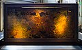
Domain: Bacteria
domain of micro-organisms .jpg) E. coli, 10 000× | |||||||
| Upload media | |||||||
| Instance of | |||||||
|---|---|---|---|---|---|---|---|
| Subclass of | |||||||
| Start time | |||||||
| Different from | |||||||
| |||||||
| |||||||
| Taxon author | Carl Woese, 2024 | ||||||
| |||||||
- Included phyla (for NCBI):
- Acidobacteria, Actinobacteria, Aquificae, Armatimonadetes, Bacteroidetes, Caldiserica, Chlamydiae, Chlorobi, Chloroflexi, Chrysiogenetes, Cyanobacteria, Deferribacteres, Deinococcus-Thermus, Dictyoglomi, Elusimicrobia, Fibrobacteres, Firmicutes, Fusobacteria, Gemmatimonadetes, Lentisphaerae, Nitrospirae, Planctomycetes, Proteobacteria, Spirochaetes, Synergistetes, Tenericutes, Thermodesulfobacteria, Thermotogae, Verrucomicrobia
- Acidobacteria, Actinobacteria, Aquificae, Bacteria incertae sedis, Bacteroidetes, Caldiserica, Chlamydiae, Chlorobi, Chloroflexi, Cyanobacteria, Deferribacteres, Deinococcus-Thermus, Elusimicrobia, Fibrobacteres, Firmicutes, Fusobacteria, Gemmatimonadetes, Lentisphaerae, Nitrospirae, Planctomycetes, Proteobacteria, Spirochaetes, Synergistetes, Tenericutes, Thermodesulfobacteria, Thermotogae, Verrucomicrobia
- Note: Only marine phyla
- Abditibacteriota, Acidobacteriota, Actinomycetota, Aquificota, Armatimonadota, Atribacterota, Bacillota, Bacteroidota, Balneolota, Caldisericota, Calditrichota, Chlamydiota, Chlorobiota, Chloroflexota, Chrysiogenota, Coprothermobacterota, Cyanobacteriota, Deferribacterota, Deinococcota, Dictyoglomerota, Elusimicrobiota, Fibrobacterota, Fusobacteriota, Gemmatimonadota, Kiritimatiellota, Lentisphaerota, Mycoplasmatota, Nitrospinota, Nitrospirota, Planctomycetota, Pseudomonadota, Rhodothermota, Spirochaetota, Synergistota, Thermodesulfobacteriota, Thermomicrobiota, Thermotogota, Verrucomicrobiota
- Note: Only valid phyla
Subcategories
This category has the following 38 subcategories, out of 38 total.
- Bacteria in Auckland Museum (18 F)
.
?
A
- Abditibacteriota (1 F)
B
C
D
- Dictyoglomi (3 F)
F
G
M
- Methylomirabilota (18 F)
N
P
R
S
T
V
Media in category "Bacteria"
The following 200 files are in this category, out of 376 total.
(previous page) (next page)-
Jer-Bactéthie.ogg 1.0 s; 12 KB
-
De-Bakterie2.ogg 1.8 s; 18 KB
-
De-Bakterium2.ogg 2.0 s; 19 KB
-
 1-s2.0-S215075112400474X-mbio.01623-24.f003 lrg.gif 1,864 × 1,097; 178 KB
-
 12864 2008 Article 1790 Fig1 HTML.png 1,184 × 486; 314 KB
-
 13068 2022 2210 Fig2 HTML.webp 1,960 × 1,438; 203 KB
-
 41396 2020 705 fig1 (cropped).jpg 1,350 × 1,521; 225 KB
-
 41396 2020 705 fig1.jpg 1,350 × 2,207; 487 KB
-
 41396 2020 705 fig1abc.jpg 1,328 × 662; 242 KB
-
 41396 2020 705 fig1d (cropped).jpg 261 × 868; 49 KB
-
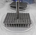 96pinner.jpg 2,810 × 2,691; 2.01 MB
-
 A bacterial colony 20200218 141234.jpg 1,200 × 1,600; 570 KB
-
 A thousands eyes (9404711386).jpg 4,928 × 3,264; 2.47 MB
-
 A. E. J. Yersin, photograph Wellcome L0016609.jpg 3,446 × 5,236; 9.29 MB
-
 Agar Art with Microbes.jpg 4,000 × 3,000; 2.27 MB
-
 Agar Plates stored in the laminar flow cabinet.jpg 3,264 × 1,836; 3.02 MB
-
 Amor por bactérias.jpg 1,269 × 1,133; 464 KB
-
 Anaerobic Chamber.pdf 1,650 × 1,275; 4.08 MB
-

-
 Antimicrobial resistance.jpg 6,720 × 4,480; 5.74 MB
-
 Aspergillus niger conidia under the microscope.jpg 2,592 × 1,936; 1.31 MB
-
 Atlas of bacterial flagellation (1960) (19721564194).jpg 1,824 × 2,828; 1.04 MB
-
 Bacillus bacteria growth under the microscope 3.jpg 5,461 × 5,461; 2.36 MB
-
 Bacteri attachment.jpg 718 × 540; 69 KB
-
 Bacteria (35323217002).jpg 1,149 × 1,564; 243 KB
-
 Bacteria (35795301244).jpg 1,149 × 1,834; 290 KB
-
 Bacteria and pus cells in a Gram stained smear of sputum.jpg 4,000 × 3,000; 1.49 MB
-
 Bacteria beginning to colonize.jpg 1,600 × 1,200; 99 KB
-
 Bacteria Bidirectional DNA Replication.png 1,082 × 903; 417 KB
-
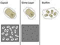 Bacteria Capsules and Slime Layers.jpg 720 × 540; 52 KB
-
 Bacteria cell wall ku.png 1,894 × 903; 523 KB
-
 Bacteria collage.jpg 2,880 × 2,160; 3.31 MB
-
 Bacteria growing in interesting structures 20200214 145246 01.jpg 6,489 × 3,286; 11.73 MB
-
 Bacteria growing in interesting structures 20200214 145246 02.jpg 4,822 × 5,545; 13.53 MB
-
 Bacteria growing in interesting structures 20200214 145246 03.jpg 4,801 × 6,006; 13.81 MB
-
 Bacteria growing in interesting structures 20200214 145246 04.jpg 6,375 × 2,867; 9.48 MB
-
 Bacteria growing in interesting structures 20200214 145246 05.jpg 3,076 × 6,157; 7.56 MB
-
 Bacteria growing in interesting structures 20200214 145246 06.jpg 4,271 × 7,009; 6.96 MB
-
 Bacteria growing in interesting structures 20200214 145246 07.jpg 2,625 × 6,782; 4.56 MB
-
 Bacteria growing in interesting structures 20200214 145246 08.jpg 5,183 × 4,413; 6.75 MB
-
 Bacteria growing in interesting structures 20200214 145246 09.jpg 7,162 × 6,909; 12.9 MB
-
 Bacteria growing in interesting structures 20200214 145246 10.jpg 6,031 × 4,346; 7.96 MB
-
 Bacteria growing in interesting structures 20200214 145246 11.jpg 5,491 × 5,219; 7.67 MB
-
 Bacteria growth on everyday objects.png 750 × 1,334; 2.42 MB
-
 Bacteria ico.png 64 × 64; 7 KB
-
 Bacteria infect tissues using ‘chemical harpoons’ (17978494003).jpg 1,100 × 790; 937 KB
-
 Bacteria involved in causing and treating cancers.svg 512 × 293; 403 KB
-
 Bacteria on everyday objects.png 750 × 1,334; 1.88 MB
-
 Bacteria planet.jpg 3,024 × 4,032; 3.92 MB
-
 Bacteria template.png 40 × 50; 3 KB
-
 Bacteria through microscope.jpg 2,448 × 3,264; 1.17 MB
-
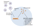 Bacteria USP response.png 406 × 311; 20 KB
-
 Bacteria yeast and fungi 01.jpg 3,264 × 2,448; 3.09 MB
-
 Bacteria yeast and fungi 02.jpg 3,264 × 2,448; 2.89 MB
-
 Bacteria yeast and fungi 03.jpg 3,264 × 2,448; 2.95 MB
-
 Bacteria yeast and fungi 04.jpg 3,264 × 2,448; 3.04 MB
-
 Bacteria yeast and fungi 05.jpg 6,016 × 4,512; 2.5 MB
-
 Bacteria yeast and fungi 06.jpg 6,016 × 4,512; 2.5 MB
-
 Bacterial and fungal colonie under the microscope 20200406 013200.jpg 7,135 × 7,346; 7.52 MB
-
 Bacterial and fungal colonie under the microscope20200406 013112.jpg 6,630 × 6,615; 7.72 MB
-
 Bacterial cell structure ku.png 1,638 × 1,065; 1.02 MB
-
 Bacterial cell walls.jpg 1,012 × 717; 795 KB
-
 Bacterial Cluster (After image enhancement).png 2,048 × 2,048; 2.35 MB
-
 Bacterial Cluster (Before image enhancement).png 2,048 × 2,048; 1.56 MB
-
 Bacterial colonies 20200214 120924 1.jpg 1,600 × 1,200; 690 KB
-
 Bacterial colonies microscope view 20200219 120541.jpg 4,769 × 4,769; 4.22 MB
-
 Bacterial colonies under the microscope 20200226 224231 01.jpg 3,676 × 5,396; 1.63 MB
-
 Bacterial colonies under the microscope 20200226 224231 02.jpg 4,429 × 4,429; 1.22 MB
-
 Bacterial colonies under the microscope20200406 013035.jpg 5,677 × 4,774; 4.13 MB
-
 Bacterial colony 20200218 145323 01.jpg 5,450 × 5,450; 3.49 MB
-
 Bacterial colony 20200218 145323 02.jpg 5,213 × 4,577; 2.98 MB
-
 Bacterial colony 20200218 145323 03.jpg 5,149 × 5,332; 3.12 MB
-
 Bacterial colony 20200218 145323 04.jpg 2,669 × 5,539; 2.09 MB
-
 Bacterial colony and fungus under the microscope 01.jpg 5,092 × 5,228; 6.25 MB
-
 Bacterial colony and fungus under the microscope 02.jpg 3,193 × 3,413; 2.59 MB
-
 Bacterial colony microscopy building interesting structures on the agar 01.jpg 3,428 × 3,342; 3.5 MB
-
 Bacterial colony microscopy building interesting structures on the agar 02.jpg 6,016 × 4,512; 3.06 MB
-
 Bacterial colony microscopy building interesting structures on the agar 03.jpg 6,539 × 6,671; 11.24 MB
-
 Bacterial colony microscopy building interesting structures on the agar 04.jpg 4,064 × 4,858; 5.14 MB
-
 Bacterial colony microscopy building interesting structures on the agar 05.jpg 6,729 × 6,676; 12.01 MB
-
 Bacterial colony microscopy building interesting structures on the agar 06.jpg 6,016 × 4,512; 3.69 MB
-
 Bacterial DNA in the gel under the UV light.jpg 1,744 × 2,125; 2.28 MB
-
 Bacterial growth under the microscope IMG 20200224 130013.jpg 5,768 × 4,550; 3.62 MB
-
 Bacterial growth under the microscope20200406 012936.jpg 5,040 × 4,740; 5.79 MB
-
 Bacterial mobile elements-fa.png 450 × 167; 36 KB
-
 Bacterial OTUs from clone libraries and next-generation sequencing.png 1,748 × 1,212; 1.3 MB
-
 Bacterial small rna mechanisms.png 1,280 × 720; 110 KB
-
 Bacterial Smear of Abiotrophia defectiva.jpg 461 × 461; 30 KB
-
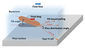 Bacterial twitching.png 1,143 × 653; 204 KB
-
 Bacterial UTI picture of urine microscopy showing plenty of pus cells and bacteria.jpg 3,264 × 2,448; 1.07 MB
-
 Bacteriapolis at the Exploratorium.jpg 4,504 × 2,772; 5.4 MB
-
 Bacterias lacticas.jpg 1,284 × 762; 73 KB
-
 Bacterias simbioticas de raíz.jpg 900 × 1,600; 133 KB
-
 Bacterias vistas en microscopio electrónico de barrido.jpg 3,072 × 2,082; 7.99 MB
-

-

-
 Bactérie acétique.jpg 1,280 × 853; 414 KB
-
 Bakterien (Bacteria) - 630x (14045255442).jpg 3,180 × 2,172; 3.02 MB
-
 Batteri.jpg 640 × 480; 22 KB
-
 BD Bactec for rapid detction of microbial growth.jpg 4,000 × 1,844; 3.94 MB
-
 Beneficial influence of plant growth-promoting rhizobacteria.png 3,469 × 2,746; 2.47 MB
-
 Beutenbergia cavernae type strain (HKI 0122T).webp 1,093 × 931; 150 KB
-
 Big Bacteria (43435845060).jpg 4,752 × 3,168; 1.11 MB
-
 Binding of SmeT to DNA.png 603 × 539; 166 KB
-
 Biomasa por peso seco 1. Dry cell weight.jpg 5,585 × 3,723; 7.88 MB
-
 Biomasa por peso seco 2. Dry cell weight.jpg 4,627 × 3,085; 4.91 MB
-
 Bison tracks in a bacteria mat at Lower Geyser Basin (54364991167).jpg 6,720 × 4,480; 10.96 MB
-
 Blood culture bottle without growth of organisms.jpg 4,000 × 3,000; 6.03 MB
-
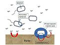
 BTEX degrading bacteria.jpg 2,304 × 4,096; 760 KB
-
 The relation of bacteria to the flavors of cheddar cheese (IA bullbai062).pdf 814 × 1,300, 37 pages; 3.39 MB
-
 C. urealyticum 2022-05-13.jpg 1,632 × 1,439; 533 KB
-
 Casein hydrolysis.png 1,856 × 2,475; 4.85 MB
-
 Cells of Kueselia aquadivae.jpg 549 × 316; 34 KB
-
 Cellular structures of a typical bacterium, planctomycetes and eukaryotes.jpg 1,762 × 688; 276 KB
-
 CEM&fungus.jpg 872 × 640; 81 KB
-

-
 Changes in swimmer velocity and Reynolds number with length scale.jpg 1,084 × 516; 291 KB
-
 Chemosynthetic Microbial Mats.jpg 3,000 × 2,250; 1.46 MB
-
 Chromatium2.jpg 750 × 304; 83 KB
-

-
 Cocobacilos.jpg 900 × 1,600; 107 KB
-
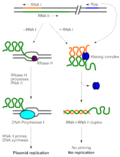 ColE1 replication control.png 4,068 × 5,324; 572 KB
-
 Colibacile pwels.jpg 234 × 256; 7 KB
-
 Colonia microbiana.jpg 640 × 480; 40 KB
-
 Colonia microbiologica de un Suelo del Alto Valle de Río Negro.jpg 3,060 × 4,080; 2.68 MB
-
 Colonias de bacterias blancas y amarillas vistas en lupa.jpg 640 × 480; 262 KB
-
 Colonie Bacillus.jpg 12,032 × 9,024; 12.82 MB
-
 Colonie bacteriana - Bacterial colony.jpg 4,857 × 5,706; 2.28 MB
-
 COLONIE BATTERICHE ISOLATE NEL TERRENO.jpg 3,870 × 2,591; 5.57 MB
-
 Colonie batteriche o vita marina?.jpg 2,505 × 2,505; 3.67 MB
-
 Colonization of potato tubers by bacteria.png 1,200 × 1,652; 1.36 MB
-
 ColorazioneCapsula.JPG 198 × 97; 2 KB
-
 Compound image from "Functional Green-Tuned Proteorhodopsin from Modern Stromatolites".png 2,150 × 1,637; 5.11 MB
-
 Conditions multiplication thermiques bacteries.png 600 × 822; 34 KB
-
 Connecting.jpg 290 × 190; 13 KB
-
 Control de Plagas.jpg 6,016 × 4,000; 14.67 MB
-

-
 Culture of Pseudoglutamicibacter cumminsii.jpg 2,848 × 2,140; 460 KB
-
 Culture Plates.jpg 4,752 × 3,168; 6.26 MB
-
 Cyanase enzymatic reaction scheme.png 1,018 × 270; 39 KB
-
 Cyanase Pentamer with labeled argenine resides.png 1,554 × 1,482; 554 KB
-
 Dark water running from a pool.jpg 3,672 × 4,896; 9.16 MB
-
 Dehalogenese durch Bakterien.jpg 620 × 414; 38 KB
-
 Dehalogenimonas alkenigignens type strain IP3-3T.png 530 × 456; 135 KB
-
 Desulfoluna.png 1,178 × 767; 1.04 MB
-
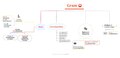
 Diagnostic algorithm of possible bacterial infection.png 5,376 × 4,133; 3.16 MB
-
 Die Gartenlaube (1879) b 063 1.jpg 1,027 × 865; 569 KB
-
 Die Gartenlaube (1891) b 362 2.jpg 252 × 301; 8 KB
-
 Die Gartenlaube (1891) b 363 1.jpg 390 × 248; 12 KB
-
 Die Gartenlaube (1891) b 363 5.jpg 275 × 279; 8 KB
-
 Die Gartenlaube (1891) b 364.jpg 118 × 326; 4 KB
-
 Die Pflanzenwelt (1913-1922.) (20913876076).jpg 2,038 × 3,218; 1.2 MB
-
 Different types of bacterial colonies.jpg 1,600 × 1,200; 748 KB
-
 Divergence from a common ancestor.png 360 × 320; 91 KB
-
 DNA barcoding of marine bacteria.jpg 500 × 129; 90 KB
-
 DNA Replication in prokaryotes.png 2,000 × 729; 408 KB
-
 Dust storms as a source of aerosolized bacteria.png 460 × 239; 122 KB
-
 E. faecalis on CLED medium.jpg 4,000 × 3,000; 1.53 MB
-
 Effect of penicillin on the coli bacteria.jpg 1,522 × 1,831; 2.94 MB
-
 Effector binding to SmeT.png 557 × 538; 313 KB
-
 El arte de lo invisible.jpg 640 × 480; 209 KB
-
 Elusimicrobium.jpg 3,479 × 1,715; 841 KB
-
 EnteroPluri Test.png 3,852 × 458; 470 KB
-
 Essential metabolic genes in bacteria.png 720 × 540; 79 KB
-
 Extreme aquatic habitats and their extremophiles and molecules.png 12,430 × 6,581; 5.26 MB
-
 Fecal flora on XLD Agar.jpg 4,000 × 3,000; 1.3 MB
-
 Fermentación de azúcares.jpg 3,896 × 3,240; 2.18 MB
-
 Fermentación mesada, microorganismos pigmentados 1A.jpg 2,605 × 3,908; 4.38 MB
-
 Fermentación mesada, microorganismos pigmentados 1B.jpg 6,000 × 4,000; 8.47 MB
-
 Fermentación mesada, microorganismos pigmentados 2A.jpg 2,655 × 3,982; 4.01 MB
-
 Fermentación mesada, microorganismos pigmentados 2B.jpg 3,125 × 4,687; 6.05 MB
-
 Fight Bac! (085 086) (7396068296).jpg 7,200 × 5,400; 6.97 MB
-
 Figure2b.pdf 1,125 × 450; 73 KB
-
 Figure2d.pdf 1,125 × 450; 100 KB
-
 Figure2e.pdf 1,125 × 450; 112 KB
-
 Flask of luminous bacteria.jpg 1,930 × 2,143; 466 KB
-
 FLoc vase Basse-Deûle rive G vers St André61.JPG 4,928 × 3,264; 1.27 MB
-

-
 Fmicb-06-00439-g005.jpg 964 × 959; 563 KB
-
 Fmicb-06-00439-g005AB-Ignicoccus-hospitalis.jpg 957 × 461; 291 KB
-
 Fmicb-06-00439-g005CD-Kuenenia-stuttgartiensis.jpg 876 × 478; 294 KB
-
 Fmicb-11-602250-g001D.jpg 549 × 879; 118 KB
-
 Fold Topology and Oligomerization States of H-NS.jpg 663 × 919; 210 KB
-
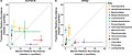 Freshwater versus marine bacterial abundance by taxonomy.jpg 1,280 × 549; 81 KB
-
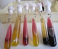
 Gas formation by bacteria in TSI agar tube.jpg 3,264 × 2,448; 1.59 MB
-
 GenericBacteriumClipArt.pdf 141 × 54; 2 KB
-
 Glucosa-lactosa-sacarosa.jpg 3,896 × 3,240; 2.19 MB
-
 Gram negative diagram.pdf 4,068 × 1,950; 234 KB
-
 Gram Positive Cocci.jpg 4,000 × 2,250; 1.23 MB
-
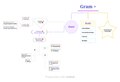 Gram positive diagram.pdf 3,375 × 2,297; 194 KB
-
 Gram's collor.jpg 3,264 × 1,836; 475 KB
-
 Grand Prismatic Spring Bacteria PLC-PK-YS-15.jpg 3,661 × 1,457; 4.61 MB
-

-
 Green running water.jpg 3,672 × 4,896; 9.09 MB
-
 Greenish bacteria and concretions.jpg 4,896 × 3,672; 8.78 MB
-
 Growth of bacteria on MHA isolated from sewage.jpg 3,000 × 4,000; 3.42 MB
-
 Growth of microbes in blood culture bottle.jpg 4,000 × 3,000; 3.71 MB
-
 Größen von Partikeln in Aerosolen.png 1,748 × 1,128; 67 KB
-
 Herminiimonas arsenitoxidans.png 889 × 697; 508 KB
-
 Host killing.png 5,391 × 3,568; 424 KB


.jpg)


.jpg)


_(19721564194).jpg)


.jpg)
.jpg)


.jpg)


.png)
.png)


_-_Planche_didactique_-_Institut_Pasteur_;_V._Roussel_(lithographe)_-_btv1b101169121.jpg)
_-_Planche_didactique_-_Institut_Pasteur_;_V._Roussel_(lithographe)_-_btv1b10116913g.jpg)

_-_630x_(14045255442).jpg)


.webp)
.jpg)


.jpg)


_b_063_1.jpg)
_b_362_2.jpg)
_b_363_1.jpg)
_b_363_5.jpg)
_(20913876076).jpg)


_(7396068296).jpg)


_in_a_BACTEC_MGIT_tube.jpg)


.jpg)
.jpg)
_b_364.jpg)